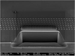
Plan view imaging of a sample surface can be performed using field emission scanning electron microscopy (FESEM), helium ion microscopy and scanning probe microscopy. Cross-section imaging can be achieved by first making a cross-sectional cut using the focussed ion beam (FIB) system and then imaging in-situ using field emission scanning electron microscopy. The FIB is also able to prepare thin samples for transmission electron microscopy and perform secondary ion mass spectroscopy (SIMS) for depth profiling of material composition. A life science SEM is available for imaging of biological samples in liquids and for the analysis of sample composition using a variety of X-ray techniques including X-ray fluorescence and X-ray computed tomography.
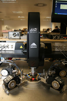
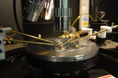
Film thicknesses can be measured using ellipsometry and step heights after etching can be measured using a profilometer. The optical properties of materials can be characterised using Raman spectroscopy and laser spectroscopy. We also have a wide range of electrical and RF testing equipment mounted on a variety of probe stations.